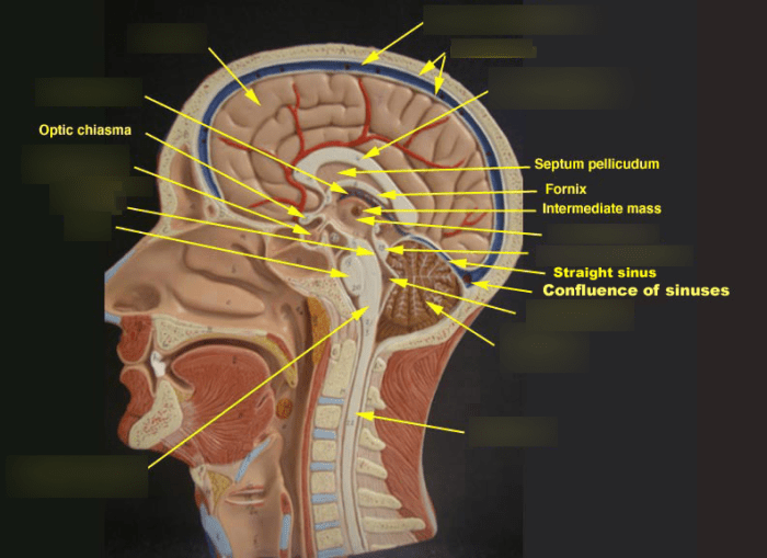
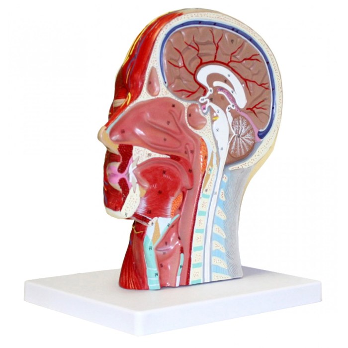
The half head with musculature labeled provides a detailed representation of the intricate network of muscles that control facial expressions and movements. This article delves into the anatomy, functions, and clinical applications of these muscles, offering a comprehensive understanding of their role in facial dynamics.
The major muscles of the half head include the frontalis, orbicularis oculi, zygomaticus major, and masseter, each with distinct functions ranging from raising eyebrows to chewing.
Half Head Anatomy

The half head, also known as the hemi-head, refers to one lateral half of the head, encompassing the skull, facial structures, and associated musculature. Understanding the anatomy of the half head is crucial for various medical and surgical procedures, including facial reconstruction and plastic surgery.
Major Muscles of the Half Head, Half head with musculature labeled
The half head musculature consists of a complex network of muscles that control facial expressions, mastication (chewing), and other functions. The major muscles of the half head include:
- Frontalis muscle:Raises the eyebrows and creates wrinkles on the forehead.
- Orbicularis oculi muscle:Closes the eyelids and controls blinking.
- Zygomaticus major muscle:Raises the corner of the mouth, creating a smile.
- Orbicularis oris muscle:Closes the lips and puckers the mouth.
- Mentalis muscle:Elevates the chin and creates a dimple.
- Buccinator muscle:Compresses the cheeks and aids in chewing.
- Masseter muscle:Elevates the mandible (lower jaw) and assists in chewing.
- Temporalis muscle:Elevates and retracts the mandible.
Muscles of Expression
The muscles of expression, also known as the mimetic muscles, play a significant role in creating facial expressions. These muscles are innervated by the facial nerve and work in combination to produce a wide range of expressions, including happiness, sadness, anger, and surprise.
For instance, the zygomaticus major muscle, along with the levator labii superioris and levator labii superioris alaeque nasi muscles, contribute to the “Duchenne smile,” a genuine expression of joy that involves raising the corners of the mouth and contracting the muscles around the eyes.
Clinical Applications
Knowledge of half head musculature is essential in various clinical settings:
- Facial injury diagnosis and treatment:Musculature labeling helps identify and treat injuries to the facial muscles and nerves.
- Reconstructive surgery:Understanding the musculature allows surgeons to restore facial function and aesthetics after trauma or disease.
Educational Resources
For further exploration of half head musculature, consider the following resources:
- Books:
- Gray’s Anatomy for Students
- Netter’s Atlas of Human Anatomy
- Articles:
- The Musculature of the Face
- Clinical Anatomy of the Facial Muscles
- Online resources:
- Visible Body Human Anatomy Atlas
- Kenhub Anatomy
User Queries: Half Head With Musculature Labeled
What are the major muscles of the half head?
The major muscles of the half head include the frontalis, orbicularis oculi, zygomaticus major, and masseter.
How do the muscles of the half head contribute to facial expressions?
The muscles of the half head work together to create a wide range of facial expressions, from smiling to frowning to raising eyebrows.
What are some clinical applications of the half head musculature?
Knowledge of the half head musculature is used in clinical settings to diagnose and treat facial injuries, as well as in reconstructive surgery.

